Why is 20 nanometers important?
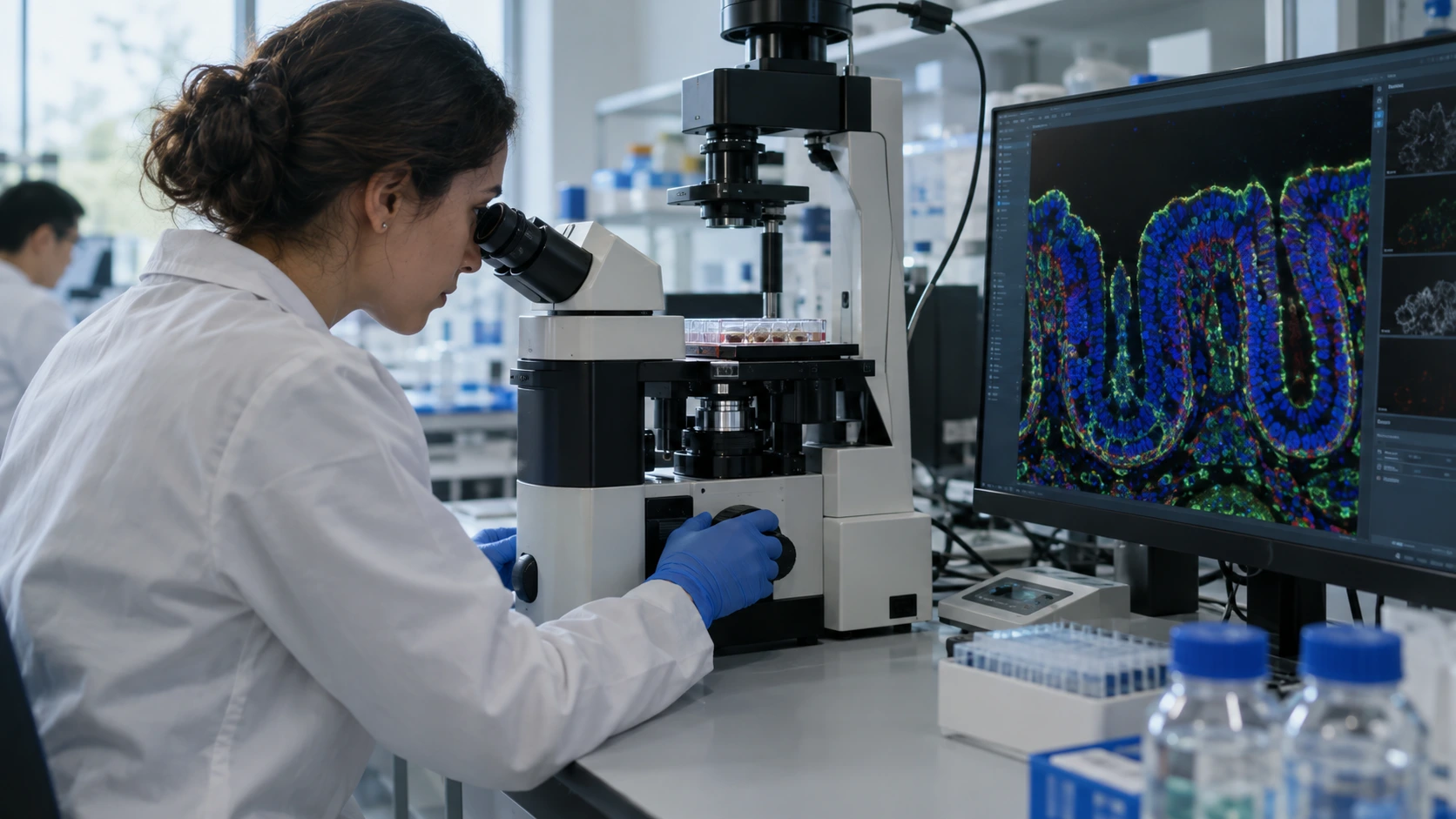
In conventional light microscopy, it is difficult to distinguish molecular structures that are very close to one another due to the diffraction limit. STED (Stimulated Emission Depletion) microscopy uses a second “depletion” laser to suppress the fluorescence signal around the focal point, creating a much smaller effective imaging area. The Rosalind Franklin Institute reports that its system can provide approximately ten-fold higher resolution, going from about 200 nanometers down to the 20-nanometer level.
What structures make up the intestinal barrier?
The intestinal epithelium forms a selective boundary between the external environment and the organism’s internal environment. Neighboring epithelial cells are organized by junctional complexes such as tight junctions, adherens junctions, desmosomes, and gap junctions. In particular, tight junction proteins play a critical role in barrier permeability by regulating which molecules can pass between cells.
What changes in IBD?
In Crohn’s disease and ulcerative colitis, genetic predisposition, immune response, microbiota, and environmental factors play a role together. Epithelial barrier dysfunction is an important part of this complex network. Alterations in tight junction architecture and permeability can be seen during inflammation; however, whether these are the initial cause of the disease or a consequence of inflammation in every patient is not always clear.
What does Curie do differently?
The Rosalind Franklin Institute’s STED platform aims not only to provide high resolution in fixed cells, but also to achieve three-dimensional imaging in thick and optically complex samples, organoids, and tissues thanks to adaptive optics. With multiplex fluorescence labeling, the relative positions of different proteins, lipids, or glycans in the same sample can be examined.
Why are patient-derived organoids valuable?
Organoids are developed by having cells obtained from a patient form three-dimensional, tissue-like structures in the laboratory. These models can display certain characteristics that are closer to real human biology than conventional two-dimensional cell cultures. By imaging tight junction organization and permeability changes in patient-derived intestinal organoids, researchers can compare the molecular architecture of different IBD phenotypes.
Barrier formation from fetal development to adulthood
Researchers from Oxford and the Rosalind Franklin Institute are conducting studies examining the development of tight junction architecture using STED in human fetal and adult intestinal tissue and paired organoids. The goal is to understand how the normal barrier is established and matures. Once a map of normal development is generated, it may be possible to see more clearly which step or protein organization has deviated in disease.
Will this technology find the “root cause” of Crohn’s?
This statement would be too strong at this stage. IBD is not a disease arising from a single cause. STED microscopy can uncover important mechanisms in barrier biology and generate new biomarkers or drug targets; however, it should not be expected to explain the entire cause of Crohn’s or ulcerative colitis on its own. The value of the technology lies in making previously optically indistinguishable molecular organization measurable.
How can it translate into new treatments?
If it can be shown that a tight junction protein is mislocalized, a cell-cell junction complex is disrupted during inflammation, or specific molecular interactions alter permeability, these mechanisms can serve as targets for drug development. However, the path from imaging findings to clinical treatment requires mechanistic validation, preclinical experiments, and human clinical trials.
Is it the same as intestinal permeability tests?
No. STED is a research imaging platform; it is not a routine test used in the clinic to diagnose “leaky gut.” Nanoscopic tight junction images and routine practical tools such as fecal calprotectin, CRP, endoscopy, histopathology, or imaging modalities answer different questions.
Our approach at our Bodrum clinic
We do not use the concept of the intestinal barrier as a label that explains every symptom on its own. In clinical evaluation, symptoms, inflammatory markers, stool tests, endoscopic-histologic findings, and, when necessary, gastroenterology evaluation should be considered together. New nanoscopy technologies deepen our understanding of the mechanism; however, as of today, they do not replace routine clinical diagnosis.
References
- The Guardian, September 14, 2026: Curie microscope and IBD research
- Rosalind Franklin Institute: STED platform, <20 nm resolution and cell junction networks
- Rosalind Franklin Institute: Tight junction research in patient-derived organoids
- UKRI, September 14, 2026: New STED microscope and IBD research investment
Last medical review: September 18, 2026